Glaucoma Screening: Early Detection for Eye Health

Glaucoma Screening: Essential for Early Detection and Vision Preservation
Glaucoma, a progressive optic neuropathy, stands as a leading cause of irreversible blindness globally, affecting over 80 million individuals worldwide. Its insidious nature often leads to significant, irreversible vision loss before symptoms are noticed, underscoring the critical importance of early detection. As Dr. Amy Catalano Boyer, a distinguished board-certified ophthalmologist and glaucoma specialist, asserts, “Proactive and accurate screening is the cornerstone of preventing vision loss from glaucoma, a condition where lost vision cannot be restored.” This comprehensive article, informed by current clinical guidelines and expert insights from specialists like Dr. Boyer, delves into the pathophysiology of glaucoma, its impact on ocular health, the essential diagnostic procedures, and the latest advancements in detection and management. Readers will gain a deeper understanding of who is at risk, evidence-based screening recommendations, and the crucial role of timely intervention in preserving visual function.
Understanding Glaucoma: Pathophysiology and Impact on Ocular Health
Glaucoma encompasses a group of chronic, progressive optic neuropathies characterized by damage to the optic nerve, typically associated with elevated intraocular pressure (IOP). This damage results in characteristic visual field defects and, if left untreated, can lead to profound and irreversible vision loss. Dr. Amy Catalano Boyer frequently highlights that “the insidious nature of glaucoma means patients often don’t notice symptoms until significant, irreversible damage to the retinal ganglion cells and optic nerve has occurred, underscoring the critical need for proactive screening.” The optic nerve, composed of millions of retinal ganglion cell axons, is vital for transmitting visual information from the retina to the brain. Its progressive degeneration leads to peripheral vision loss, which can advance to central vision impairment and ultimately, complete blindness. A thorough understanding of glaucoma’s pathophysiology and its impact on ocular health is paramount for effective prevention and management.
Types and Etiology of Glaucoma
Glaucoma is broadly categorized into several types, with primary open-angle glaucoma (POAG) being the most prevalent, accounting for approximately 90% of cases in Western populations. POAG is characterized by an open, normal-appearing anterior chamber angle, but with impaired aqueous humor outflow through the trabecular meshwork, leading to elevated IOP. Angle-closure glaucoma (ACG), less common but often more acute, occurs when the iris obstructs the drainage angle, impeding aqueous humor outflow. Secondary glaucomas can arise from other ocular or systemic conditions, such as trauma, inflammation, certain medications (e.g., corticosteroids), or pre-existing medical conditions like diabetes and hypertension. Genetic predisposition, advanced age, and specific ethnic backgrounds are significant risk factors. Recognizing these distinct types and their underlying etiologies is crucial for accurate diagnosis and targeted management.
Mechanism of Optic Nerve Damage in Glaucoma
The primary mechanism of optic nerve damage in glaucoma involves the progressive apoptosis (programmed cell death) of retinal ganglion cells and their axons. While elevated IOP is a major risk factor, it is not the sole determinant; other factors such as vascular dysregulation, oxidative stress, and neuroinflammation also contribute. Elevated IOP mechanically compresses the optic nerve fibers at the lamina cribrosa, disrupting axonal transport and blood supply. This sustained stress leads to the degeneration of nerve fibers, which manifest as thinning of the retinal nerve fiber layer (RNFL) and characteristic changes to the optic disc, such as increased cup-to-disc ratio and neuroretinal rim thinning. Dr. Amy Catalano Boyer explains, “The optic nerve is akin to the main data cable connecting your eye to your brain. Once those delicate nerve fibers are damaged, they do not regenerate, which is why early detection before extensive axonal loss is paramount to preserving functional vision.” The irreversible nature of this damage underscores the urgency of early diagnosis and intervention.
Essential Glaucoma Diagnostic Tests for Early Detection

Early and accurate detection of glaucoma is paramount for effective disease management and the preservation of vision. Dr. Amy Catalano Boyer, a leading expert in glaucoma screening, emphasizes that “a comprehensive ophthalmic examination utilizing a multi-modal diagnostic approach is indispensable for accurately identifying glaucoma in its earliest, often asymptomatic, stages.” Several diagnostic tests are essential for identifying the condition before significant and irreversible optic nerve damage occurs.
Intraocular Pressure Measurement (Tonometry)
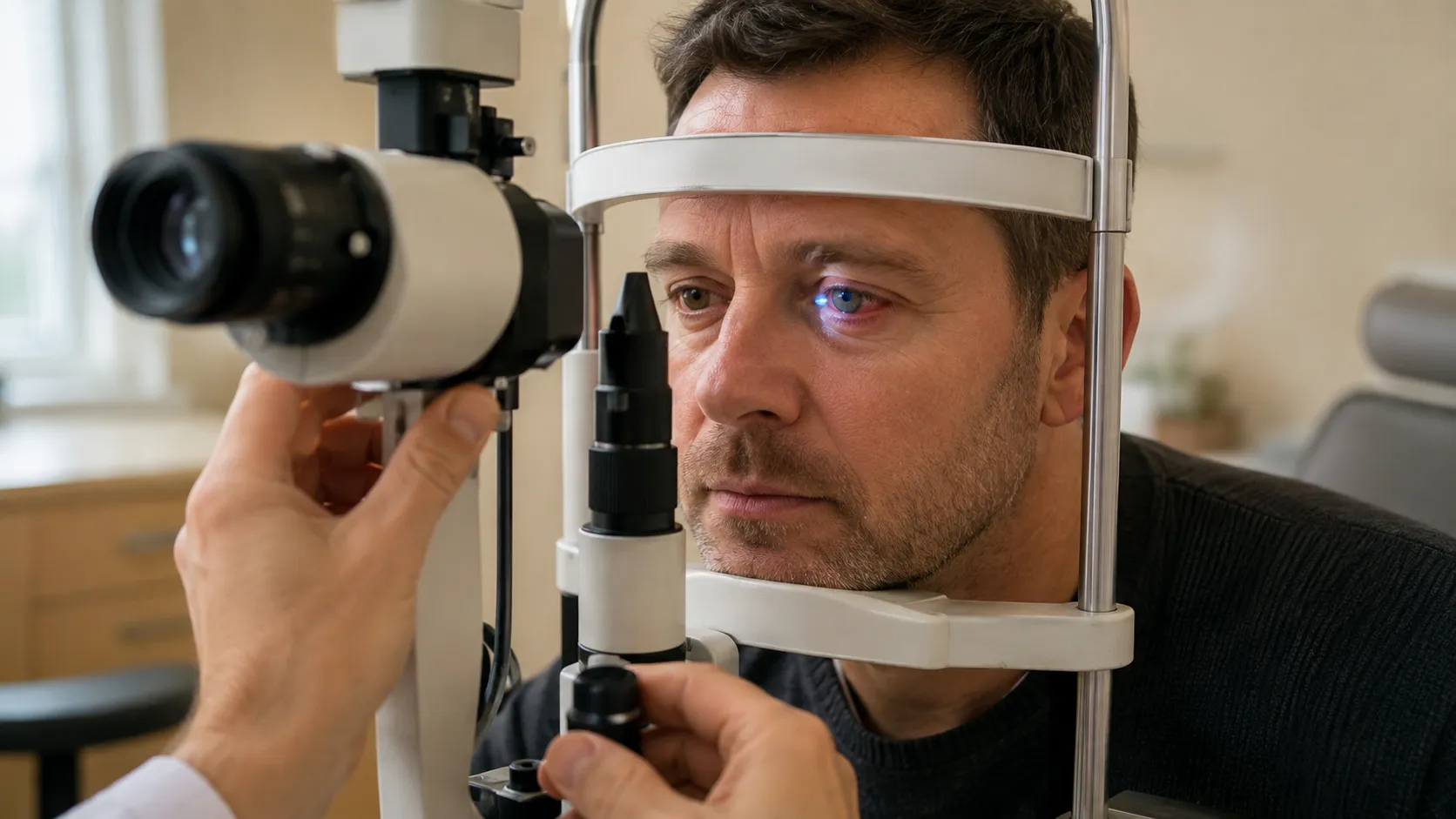
Tonometry is a fundamental test that measures the intraocular pressure (IOP) within the eye. Elevated IOP is the most significant modifiable risk factor for glaucoma. The gold standard for IOP measurement is Goldmann Applanation Tonometry, which measures the force required to flatten a standard area of the cornea. Other methods include non-contact tonometry and rebound tonometry. Dr. Boyer notes, “While an elevated IOP reading is a critical indicator, it is not solely diagnostic of glaucoma. We interpret IOP within the context of other clinical findings, but tonometry often provides our initial clue and guides further investigation.” Normal IOP typically ranges from 10-21 mmHg, but glaucoma can occur at any pressure (normal-tension glaucoma), and elevated IOP does not always lead to glaucoma (ocular hypertension). Therefore, regular IOP checks are vital, especially for individuals with other risk factors.
Ophthalmoscopy, Gonioscopy, and Visual Field Testing
These three tests are complementary and crucial for a comprehensive glaucoma assessment:
- Ophthalmoscopy: This involves a stereoscopic examination of the optic nerve head to assess its morphology. Key indicators of glaucomatous damage include an increased cup-to-disc ratio, thinning or notching of the neuroretinal rim, disc hemorrhages, and asymmetry between the two eyes. Direct and indirect ophthalmoscopy allow the clinician to visualize these subtle changes.
- Gonioscopy: This diagnostic procedure uses a specialized lens to directly visualize the anterior chamber angle, where the iris meets the cornea. It is essential for classifying glaucoma as open-angle or angle-closure, which dictates appropriate treatment strategies.
- Visual Field Testing (Perimetry): This functional test assesses the integrity of the visual pathway by mapping the patient’s peripheral and central vision. The Humphrey Visual Field Analyzer is commonly used to detect characteristic patterns of glaucomatous vision loss, such as nasal steps, arcuate scotomas, and paracentral defects. These defects often correlate with structural damage to the optic nerve.
Dr. Amy Catalano Boyer explains, “Ophthalmoscopy provides us with a direct, structural view of the optic nerve’s health, gonioscopy clarifies the drainage angle anatomy, and visual field tests reveal how well a patient is actually seeing and if functional vision loss has occurred. All three are indispensable for a complete and accurate glaucoma assessment.” Together, these diagnostic procedures form a robust, evidence-based approach to early detection and monitoring.
Glaucoma Screening: Who, When, and Evidence-Based Guidelines
Understanding who should be screened for glaucoma and the appropriate timing is essential for effective prevention and early intervention. Dr. Amy Catalano Boyer frequently advises her patients on personalized screening schedules, emphasizing the importance of proactive, risk-stratified care based on established clinical guidelines.
Key Risk Factors for Glaucoma
Several well-established risk factors significantly increase an individual’s likelihood of developing glaucoma. These include:
- Age: The prevalence of glaucoma increases significantly with age, particularly after 60.
- Family History: A first-degree relative with glaucoma increases an individual’s risk by 5-9 times.
- Ethnicity: Individuals of African descent have a 4-5 times higher risk of developing POAG, often at an earlier age and with more severe progression. Hispanic individuals also have an increased risk.
- Elevated Intraocular Pressure (IOP): While not diagnostic, persistently high IOP is the strongest modifiable risk factor.
- Central Corneal Thickness (CCT): Thinner corneas are associated with a higher risk of developing glaucoma and may lead to underestimation of true IOP.
- Systemic Diseases: Diabetes, hypertension, and cardiovascular disease are associated with an increased risk.
- High Myopia: Severe nearsightedness is a risk factor for POAG.
- Corticosteroid Use: Prolonged use of corticosteroids (topical, oral, inhaled) can elevate IOP.
Dr. Amy Catalano Boyer stresses, “If you possess one or more of these risk factors, particularly a strong family history, your risk is significantly elevated, making regular, early, and comprehensive screenings non-negotiable.” Recognizing these risk factors empowers individuals to seek appropriate ophthalmic care proactively.
Understanding these risk factors is further supported by comprehensive research into the epidemiology and screening guidelines for open-angle glaucoma.
Glaucoma Risk Factors & Screening Guidelines
Open-angle glaucomas are a group of chronic progressive optic nerve neuropathies with a gonioscopic open anterior chamber angle. They are one of the main causes of visual impairment and blindness in industrialized countries. The aim of this article is to discuss and evaluate the epidemiology and risk factors for the development of open-angle glaucoma and to present the screening procedure for open-angle glaucoma according to the recently published S2e guidelines of the Association of the Scientific Medical Societies in Germany (AWMF).
Risk factors for open-angle glaucoma and recommendations for glaucoma screening, AK Schuster, 2021
For broader clinical guidelines and professional resources on eye care, the American Academy of Ophthalmology provides extensive information.
Recommended Age and Frequency for Glaucoma Screening
Screening recommendations are stratified based on age and the presence of risk factors, as outlined by professional organizations like the American Academy of Ophthalmology (AAO):
- Under 40 with no risk factors: Comprehensive eye exam every 5-10 years.
- Ages 40-54 with no risk factors: Comprehensive eye exam every 2-4 years.
- Ages 55-64 with no risk factors: Comprehensive eye exam every 1-3 years.
- Age 65 and older with no risk factors: Comprehensive eye exam every 1-2 years.
- Individuals with any risk factors (e.g., family history, African descent, diabetes): Screenings should begin earlier and occur more frequently, often annually or every 6-12 months, regardless of age.
Dr. Amy Catalano Boyer advises, “While a baseline comprehensive eye exam around age 40 is a prudent starting point for everyone, those with significant risk factors should initiate screenings earlier and adhere to more frequent follow-up schedules, often annually or even semi-annually, as determined by their ophthalmologist.” Establishing and adhering to a personalized screening schedule is crucial for the timely detection and management of glaucoma, significantly improving the prognosis for vision preservation.
Lifestyle and Preventative Measures Supporting Ocular Health
While lifestyle choices cannot prevent glaucoma in individuals predisposed to the condition, they play a significant supportive role in maintaining overall ocular health and may help mitigate risk factors or complement medical treatments in managing glaucoma progression. Dr. Amy Catalano Boyer often discusses with her patients how daily habits can contribute to preserving vision and overall well-being.
Dietary and Exercise Habits for Ocular Health
A balanced diet rich in specific nutrients can support ocular health. Key components include:
- Antioxidants: Found in leafy green vegetables (spinach, kale), berries, and colorful fruits, antioxidants like lutein, zeaxanthin, and anthocyanins help protect retinal cells from oxidative stress.
- Omega-3 Fatty Acids: Present in fatty fish (salmon, tuna), flaxseeds, and walnuts, omega-3s have anti-inflammatory properties beneficial for overall eye health.
- Vitamins: Vitamins A, C, and E, along with zinc, are crucial for maintaining healthy vision and protecting against various eye conditions.
Regular moderate-intensity aerobic exercise, such as brisk walking or cycling, has been shown to temporarily lower IOP and improve ocular blood flow. Dr. Boyer notes, “While diet and exercise are not a cure for glaucoma, they are powerful allies in supporting overall ocular health, managing systemic risk factors like hypertension and diabetes, and can play a supportive role in managing glaucoma progression.” Incorporating these dietary and exercise habits is a proactive approach to enhancing eye health.
Lifestyle Modifications for Glaucoma Protection

In addition to diet and exercise, several lifestyle modifications can contribute to protecting against glaucoma and its progression:
- Smoking Cessation: Smoking is a significant risk factor for many chronic diseases, including those that can impact eye health and potentially exacerbate glaucoma.
- Blood Pressure Management: Maintaining healthy blood pressure is crucial, as both high and low blood pressure can affect ocular blood flow and optic nerve health.
- Stress Management: Chronic stress can have systemic effects that may indirectly impact ocular health. Techniques like meditation and yoga can be beneficial.
- UV Protection: Wearing sunglasses that block 99-100% of UVA and UVB rays helps protect ocular tissues from harmful radiation.
- Limiting Caffeine Intake: While not definitively linked, excessive caffeine intake may temporarily elevate IOP in some individuals.
- Hydration: Maintaining adequate hydration is important for overall physiological function.
Adopting these comprehensive lifestyle practices can enhance overall well-being and contribute to a reduced risk of glaucoma progression, complementing medical management strategies.
Latest Advances in Glaucoma Screening and Monitoring
Technological advancements are continuously revolutionizing glaucoma screening and monitoring methods, significantly enhancing early detection capabilities and precision in tracking disease progression. Dr. Amy Catalano Boyer is at the forefront of integrating these innovations to provide the most advanced and personalized care for her patients.
Optical Coherence Tomography (OCT) for Early Detection
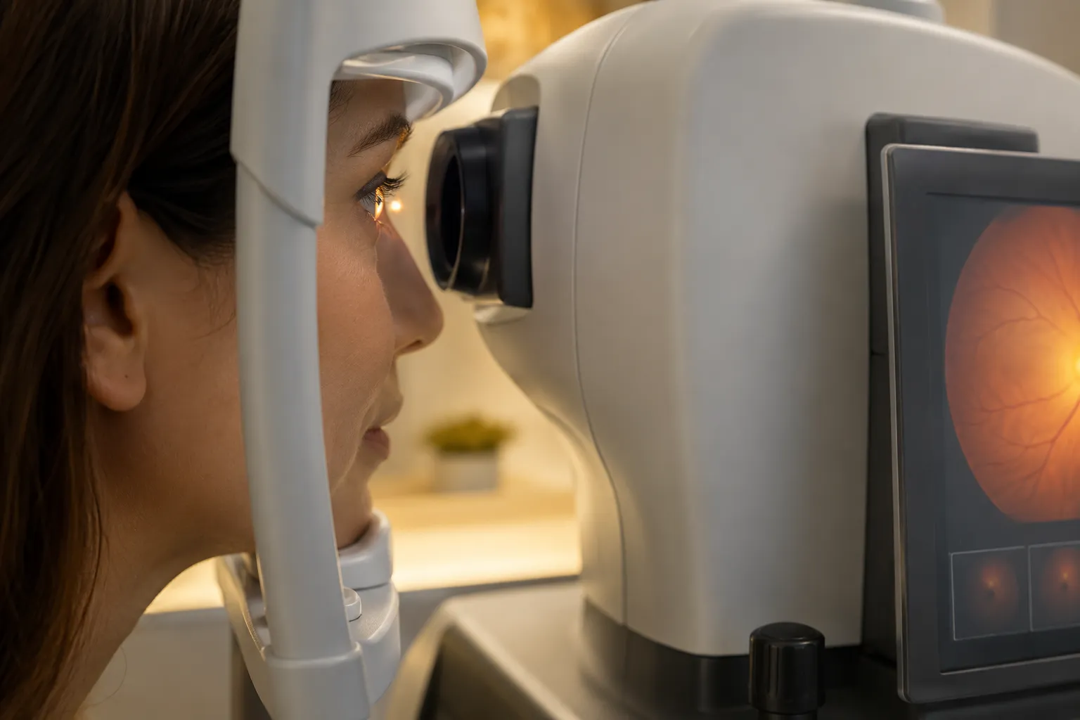
Optical Coherence Tomography (OCT) is a non-invasive, high-resolution imaging technique that provides cross-sectional images of ocular structures, particularly the optic nerve head, retinal nerve fiber layer (RNFL), and macular ganglion cell complex (GCC). OCT utilizes light waves to generate detailed anatomical maps, allowing for quantitative measurement of RNFL thickness and GCC density. These measurements are critical because glaucomatous damage often manifests as thinning in these layers, even before functional visual field defects are detectable. Dr. Amy Catalano Boyer frequently relies on OCT, stating, “OCT provides us with incredibly precise, micron-level views of the optic nerve and retina, allowing us to detect subtle structural changes indicative of glaucoma even before functional vision loss occurs. It’s an indispensable tool for early diagnosis, differentiating glaucoma from other optic neuropathies, and monitoring progression over time.” OCT has become an invaluable tool in modern ophthalmology, significantly improving diagnostic accuracy and enabling earlier intervention.
Further emphasizing the importance of advanced imaging, research highlights OCT and OCTA as crucial for early detection and monitoring of glaucoma progression.
OCT & OCTA: Essential Tools for Glaucoma Detection
Optical coherence tomography and optical coherence tomography angiography: essential tools for detecting glaucoma and disease progression, Y Shiga, 2023
Emerging Diagnostic Methods for Glaucoma
The field of glaucoma diagnostics is rapidly evolving with several promising emerging methods:
- Optical Coherence Tomography Angiography (OCTA): This advanced OCT variant non-invasively visualizes the microvasculature of the retina and optic nerve head. Glaucoma is associated with reduced blood flow, and OCTA can detect subtle changes in vessel density, potentially offering an earlier biomarker for disease onset and progression.
- Artificial Intelligence (AI) and Machine Learning: AI algorithms are being developed to analyze vast amounts of clinical data, including OCT scans, visual fields, and fundus photographs, to identify subtle patterns indicative of glaucoma. These systems can assist in risk stratification, diagnosis, and prediction of progression with high accuracy.
- Genetic Testing: Research is ongoing to identify genetic markers associated with glaucoma susceptibility and progression. While not yet routine, genetic testing may eventually help personalize screening and treatment strategies.
- Advanced Functional Testing: Beyond standard perimetry, new functional tests like frequency doubling technology (FDT) perimetry and short-wavelength automated perimetry (SWAP) are designed to detect visual field defects earlier by targeting specific retinal ganglion cell populations.
Dr. Amy Catalano Boyer actively participates in research and clinical trials to integrate these innovations, noting, “The future of glaucoma care lies in personalized medicine, driven by these advanced diagnostic tools that allow us to predict progression, customize treatments, and monitor efficacy with unprecedented accuracy and sensitivity.” Staying informed about these advancements is essential for both healthcare providers and patients to leverage the most effective strategies for vision preservation.
Glaucoma Management After Diagnosis: Treatment and Monitoring
Once a diagnosis of glaucoma is established, effective and individualized management strategies are crucial for controlling the disease and preserving remaining vision. Dr. Amy Catalano Boyer works closely with her patients to develop comprehensive treatment plans, emphasizing ongoing monitoring, patient education, and adherence to therapy.
Available Treatment Options for Glaucoma
The primary goal of glaucoma treatment is to lower intraocular pressure (IOP) to a target level that prevents further optic nerve damage. Treatment options typically include:
- Topical Medications (Eye Drops): These are often the first line of defense. Classes include:
Prostaglandin Analogs (e.g., latanoprost): Increase aqueous humor outflow.Beta-Blockers (e.g., timolol): Decrease aqueous humor production.Alpha-Adrenergic Agonists (e.g., brimonidine): Decrease aqueous production and increase outflow.Carbonic Anhydrase Inhibitors (e.g., dorzolamide): Decrease aqueous humor production.Rho Kinase Inhibitors (e.g., netarsudil): Increase aqueous outflow through the trabecular meshwork. - Laser Therapy:Selective Laser Trabeculoplasty (SLT): A non-invasive procedure that uses a low-energy laser to improve aqueous humor outflow through the trabecular meshwork, typically for open-angle glaucoma.Argon Laser Trabeculoplasty (ALT): Similar to SLT but uses a different laser wavelength.Laser Peripheral Iridotomy (LPI): Creates a small opening in the iris to prevent or treat angle-closure glaucoma.
- Surgical Interventions: Reserved for cases where medications and laser therapy are insufficient or poorly tolerated.
Trabeculectomy: Creates a new drainage pathway for aqueous humor, forming a “bleb” under the eyelid.Glaucoma Drainage Devices (e.g., Ahmed, Baerveldt implants): Involve implanting a small tube with a plate to drain aqueous humor to a reservoir under the conjunctiva.Minimally Invasive Glaucoma Surgery (MIGS): A group of procedures designed to lower IOP with less tissue trauma and faster recovery, often performed in conjunction with cataract surgery. Examples include iStent, Hydrus Microstent, and Xen Gel Stent.
Dr. Boyer explains, “Our primary objective is to lower intraocular pressure to a level that effectively prevents further optic nerve damage, individualized for each patient’s disease severity and progression rate. We typically initiate treatment with the least invasive options and escalate as needed, always prioritizing the patient’s long-term visual function and quality of life.” Understanding these diverse options is vital for patients to make informed decisions in collaboration with their ophthalmologist.
Monitoring Glaucoma Progression Over Time
Effective glaucoma management necessitates vigilant and consistent monitoring of disease progression. This involves regular, scheduled follow-up examinations that include:
- Serial IOP Measurements: Tracking IOP fluctuations and ensuring it remains within the individualized target range.
- Optic Nerve Head and RNFL Assessment: Repeated stereoscopic optic disc examinations and serial OCT imaging to detect subtle structural changes and thinning over time.
- Visual Field Testing: Regular perimetry to identify new visual field defects or progression of existing ones. Trend analysis of visual field data is crucial.
- Gonioscopy: Periodically repeated to assess the anterior chamber angle, especially if there are changes in IOP or symptoms.
- Pachymetry: Measurement of central corneal thickness, which can influence IOP readings and is a risk factor for progression.
Dr. Amy Catalano Boyer stresses the paramount importance of consistent follow-up, stating, “Glaucoma is a lifelong, chronic condition that demands vigilant, systematic monitoring. Regular check-ups allow us to track disease progression, adjust treatments promptly, and ultimately preserve our patients’ sight and quality of life by preventing further irreversible damage.” Consistent, evidence-based monitoring is essential for managing the condition effectively and preventing vision loss.
This table summarizes essential glaucoma diagnostic and monitoring tests, their clinical purposes, and recommended frequencies, highlighting the importance of a multi-modal, regular approach to managing ocular health and preventing vision loss.
Glaucoma screening is an indispensable component of maintaining ocular health and preventing irreversible vision loss. As Dr. Amy Catalano Boyer, a leading authority in ophthalmology and glaucoma care, consistently advocates, “Empowering patients with accurate knowledge, facilitating access to early, precise screening, and ensuring adherence to evidence-based management strategies are our most potent defenses against glaucoma’s devastating effects.” By understanding the complex pathophysiology of glaucoma, recognizing personal risk factors, adhering to recommended screening schedules, and staying informed about the latest advancements in diagnostic and therapeutic modalities, individuals can take proactive and informed steps to protect their vision. Regular, comprehensive ophthalmic examinations, coupled with appropriate lifestyle modifications and expert medical guidance, are essential for effective glaucoma management, a journey best navigated with the specialized expertise provided by professionals like Dr. Boyer.